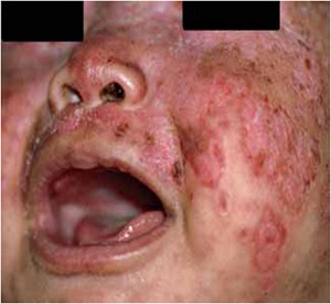
بیمار شیرخوار 6 ماهه ای است که از دو ماهگی دچارپاپول و پلاکهای اریتماتو مختصر پوسته دار روی صورت به خصوص پیشانی و اطراف چشمها شده که به تدریج گردن،تنه و اندامها را گرفتار کرده است.حال عمومی وی خوب بوده و مشکلی در بدو تولد نداشته است.
DX or DDX?
مادر بیمار سابقه درد مفاصل دست را از حدود سه سال قبل ذکر می کرد ولی تشخیص خاصی داده نشده بود.در نهایت با بررسی های آزمایشگاهی و بیوپسی پوست تشخیص بیماری شیرخوار داده شد.بیماری مادر وی نیز تشخیص داده شد.
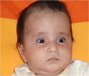
Erythematous,�annular lesions with�well-defined margins�and lighter-colored�centers on the face�and periocular�regions
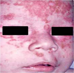
Erythematous, squamous, slightly atrophic�lesions on the face, with polycyclic margins
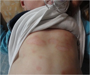
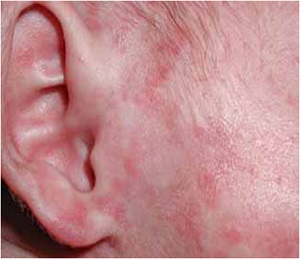
Annular lesions with erythematous, raised margins,�with a lighter-colored center, located in the preauricular region
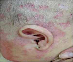
Annular, erythematous,�slightly atrophic�plaques with telangiectasia�on the scalp, in�the temporal-occipital�region and neck
Neonatal lupus erythematosus
Neonatal lupus erythematosus (NLE) is characterized by the presence of skin lesions, abnormal cardiac function or both in newborn infants of mothers with auto-antibodies against Ro, La and/or, less commonly, U1RNP
It is a rare disease that affects approximately 1 in every 20,000 live-born infants, principally girls (65%)
It presents in the children of women who have been diagnosed with systemic lupus erythematosus (SLE), Sjogren’s syndrome and other types of collagen-vascular diseases
Approximately 50% of mothers are asymptomatic at the time of diagnosis, testing positive only against Ro auto-antibodies
It presents in the children of women who have been diagnosed with systemic lupus erythematosus (SLE), Sjogren’s syndrome and other types of collagen-vascular diseases
Approximately 50% of mothers are asymptomatic at the time of diagnosis, testing positive only against Ro auto-antibodies
The pathogenesis of this disorder remains to be fully clarified; however, the transplacental passage of antibodies, principally anti-Ro antibodies (present in 95% of cases of children with NLE and their mothers) is fundamental
The transitory nature of the manifestations that coincides with the disappearance of the antibodies circulating in the infant’s blood tends to confirm this hypothesis
The Ro antigen is found in the skin, heart, liver, bowel, lungs, brain and blood cells, the tissues that are most often affected by NLE.
Ultraviolet light and estrogens increase Ro antigen expression on the surface of the keratinocyte
Nevertheless, the presence of autoantibodies is necessary but insufficient in itself to produce NLE.
There is even discordance between twins; therefore, it is believed that unknown factors exist that render some infants susceptible in the presence of maternal autoantibodies
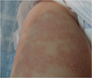
The skin lesions provoked by NLE may be congenital (20%) or appear during the first three months of life. They consist of erythematous, squamous plaques with raised margins and lighter colored centers.
They are annular or polycyclic in shape and disappear before the infant completes one year of life.
In the majority of patients, the lesions appear in photoexposed areas of the body (the face and scalp), although the skin of any other region may also be affected
Periocular erythema, referred to as “raccoon eyes” is a very common characteristic. Purplish, atrophic telangiectasias and erythema multiform-like lesions are less common
Treatment of the skin lesions consists of photoprotection and low-strength topical corticosteroids. Laser treatment may be necessary for residual telangiectasias.
The cardiologic abnormality that is characteristic of NLE is third-degree atrioventricular block, which develops in the second trimester of pregnancy and is permanent
Fetal brachycardia raises the suspicion of NLE during pregnancy and may be confirmed by fetal echocardiography.
The mortality rate in infants with congenital heart block (CHB) is around 20% even with implantation of a pacemaker, a procedure that is required in 50-100% of cases.
The risk of heart involvement in the firstborn child of an anti-Ro-positive mother is 1-2% and occurrence of the same disorder in a second child is 18%.
The efficacy of intravenous immunoglobulin is currently being evaluated for the prophylactic treatment of CHB in high-risk pregnancies (mothers who already have children with NLE) and it is recommended that fetal echocardiography should be performed from the sixteenth week of pregnancy onwards in all anti-Ropositive mothers
Liver involvement in NLE, which occurs in 10% of cases, usually manifests as liver failure, cholestasis or an increase in transaminase levels, which resolved spontaneously.
The most commonly described hematological manifestations (10% of cases) are thrombocytopenia, anemia, neutropenia and pancytopenia. These alterations are usually transitory and mild
Other organs such as the lungs and digestive tract are rarely affected.
The possibility of neurological involvement associated with NLE has been investigated.
Studies were conducted in which patients with NLE or the children of anti-Ro-positive mothers, were evaluated by neuroimaging and cephalic perimeter measurement. A greater frequency of abnormalities was found in these patients compared to healthy controls.
Differential diagnoses of skin lesions are made with seborrheic dermatitis, tinea corporis, psoriasis, atopic dermatitis, granuloma annulare, neonatal acne, erythema multiforme, Langerhans cell histiocytosis and congenital infections
The diagnosis of NLE is established from characteristic clinical findings and the identification of the abovementioned antibodies in the mother and child.
Histopathology of the skin is compatible with subacute cutaneous lupus, although in general this is not necessary for confirmation of diagnosis
The prognosis of the disease is defined by the cardiac involvement, since most of the other manifestations are transitory and self-resolving.
Long-term follow-up is vital in these patients, since they are at an increased risk of developing autoimmune diseases in late childhood or adulthood.

